

Resources and Information on Axolotls
This is catalogue of gathered articles, studies, and publishings that focus on everything relating to Ambystoma mexicanum. Axolotls have a very long, complex, and fascinating role in science, culturally, environmentally, and within the pet trade. These impactful animals have made a splash in various aspect of scientific research, primarily for their regenerative capabilities, but their unusual appearance has also captured the interest of millions of aquarium keepers worldwide for centuries!

Pudge, an adult female wild type axolotl. Credit to @smashleyyyy on Discord.
Histories of Ambystoma mexicanum
"The Mexican axolotl (Ambystoma mexicanum) is an icon of culture, a revered aquarium pet, and a highly valued animal model in biomedical research. Unfortunately, Mexican axolotls are critically endangered in their natural Xochimilco habitat in Mexico City. If axolotls go extinct, current efforts to conserve the Xochimilico ecosystem will be undermined, as will efforts to genetically manage the laboratory populations that are needed to sustain research efforts around the world. A concerted global effort is needed to protect and manage this irreplaceable species in natural and laboratory environments."
"Ambystoma mexicanum (axolotl) embryos and juveniles have been used as model organisms for developmental and regenerative research for many years. This neotenic aquatic species maintains the unique capability to regenerate most, if not all, of its tissues well into adulthood. With large externally developing embryos, axolotls were one of the original model species for developmental biology... Here, we provide an updated axolotl-staging chart ranging from one-cell stage to immature adult, paired with a perspective on both historical and current axolotl research that spans from their use in early studies of development to the recent cutting-edge research, employment of transgenesis, high-resolution imaging, and study of mechanisms deployed in regeneration."
"Since 1245 up to the mid-twentieth century, when urban sprawl drove the axolotl to near-extinction, dwellers of the Basin of Mexico have eaten axolotls. (This Nahuatl word is pronounced ah-SHO-lotl). What this chapter explores is the possibility that by 1200 BC the Early Formative inhabitants of Lake Chalco also exploited the axolotl, probably for food. It suggests that the Formative Period dwellers of the Basin of Mexico devised a graphic symbol for the axolotl – in the form of its branchlike gill stalks. As the following study explains, the symbol may have referred to the axolotl as a food. It also may have signified the regenerative ability of this perpetually embryonic creature or the mistaken notion that the aquatic axolotl can transform into a terrestrial salamander. But before considering the early prehistory of the axolotl, the chapter looks at its “history” – what has been written about it since 1521 including its enormous bibliography in today’s scientific literature – and its role in Aztec mythology."
A transcribed entry by M. Auguste Duméril in 1866, which described the rudimentary experiments that he preformed, and observations of the captive axolotls that were imported from Mexico for the first time. His research sparked centuries of scientific research on regeneration and amphibians.
"Today the Mexican axolotl is critically endangered in its natural habitat in lakes around Mexico City, but thrives in research laboratories around the world, where it is used for research on development, regeneration, and evolution. Here, we concentrate on the early history of the axolotl as a laboratory animal to celebrate that the first living axolotls arrived in Paris in 1864, 150 years ago. Maybe surprisingly, at first the axolotl was distributed across Europe without being tied to specific research questions, and amateurs engaged in acclimatization and aquarium movements played an important role for the rapid proliferation of the axolotl across the continent. But the aquarium also became an important part of the newly established laboratory, where more and more biological and medical research now took place. Early scientific interest focused on the anatomical peculiarities of the axolotl, its rare metamorphosis, and whether it was a larva or an adult... Nowadays, technical developments such as transgenic lines, and the very strong interest in stem cell and regeneration research has again catapulted the axolotl into becoming an important laboratory animal."
"The salamander Ambystoma mexicanum, commonly called “the axolotl” has a long, illustrious history as a model organism, perhaps with one of the longest track records as a laboratory-bred vertebrate, yet it also holds a prominent place among the emerging model organisms. Or rather it is by now an “emerged” model organism, boasting a full cohort molecular genetic tools that allows an expanding community of researchers in the field to explore the remarkable traits of this animal including regeneration, at cellular and molecular precision—which had been a dream for researchers over the years. This chapter describes the journey to this status, that could be helpful for those developing their respective animal or plant models."
"One of many research questions investigated in the axolotl is regeneration. The species' astonishing ability to regenerate tissues and entire body parts already became apparent shortly after the first 34 living axolotls had been brought from Mexico to Europe in 1864. In the context of their unclear status as larvae or adults and the mysterious transformation of some animals into an adult form, the Paris zoologist Auguste Duméril cut off the gills of several individuals in an attempt to artificially induce the metamorphosis. This produced the first reports on the animals' regenerative powers and led to sporadic but continuous investigations. But it remained just one of the many phenomena studied in axolotls..."

Cosmo, a juvenile melanoid leucistic axolotl.Credit to @russellmmo on Discord.
A Closer Look at Axolotls
"The critical water quality parameters that directly affect the axolotl's health include water temperature, ammonia (NH3), nitrite (NO2-), nitrate (NO3-), pH, carbonate hardness (KH, also known as alkalinity), general hardness (GH, also known as permanent hardness) and dissolved oxygen (DO). Additionally, some parameters have diurnal fluctuations. Imbalances in any one of these parameters may have an impact on others and these inter-relationships will be explained. But before the specifics of each water quality parameter is discussed, they must understand the nitrogen cycle."
"This study evaluates the viability of restored and artificial wetlands for axolotl conservation by comparing movement patterns, home range sizes, and habitat use. Using VHF telemetry, we tracked captive-bred axolotls released into both environments. Axolotls survived and foraged successfully in both sites... Recaptured individuals gained weight, suggesting successful adaptation, although two axolotls were lost to avian predation in Xochimilco after the study concluded. These findings highlight the potential of artificial wetlands like La Cantera Oriente for axolotl conservation by providing stable conditions that may mitigate habitat degradation and climate change impacts. The study recommends integrating native and artificial habitats into conservation strategies, incorporating predator awareness training before release, and ongoing habitat monitoring to enhance survival outcomes for this iconic species."
"The axolotl is a key model to study appendicular regeneration. The limb complexity resembles that of humans in structure and tissue components; however, axolotl limbs develop postembryonically. In this work, we evaluated the postembryonic development of the appendicular skeleton and its changes with aging… In axolotls, bone maturation is a continuous process that extends throughout their life. Ossification of the appendicular bones is slow and continues until the complete element is ossified. The cellular components of the appendicular skeleton change accordingly during ossification, creating a heterogenous landscape in each element. The continuous maturation of the bone is accompanied by a continuous body growth."
"... In addition, we have recently shown that a population of UV photoreceptors is also present in the retina of the neotenic Mexican salamander Ambystoma mexicanum. In this article we have presented a summary of our experiments as well a discussion of their implications for research concerning the visual system of urodeles. We have attempted to present a simplified version of our techniques so that those unfamiliar with visual physiology can gain an understanding of the experiments."
"The Axolotl, Ambystoma mexicanum, endemic to the freshwater lakes, Xochimilco and Chalco in Mexico City, feeds on zooplankton during its larval stages. We evaluated the functional response over eight weeks of A. mexicanum fed different prey items found in its natural habitat (rotifers: Brachionus havanaensis, B. calyciflorus, B. rubens and Plationus patulus; cladocerans: Moina macrocopa, Macrothrix triserialis, Alona glabra and Simocephalus vetulus; and ostracods: Heterocypris incongruens). Zooplankton consumption by A. mexicanum varied in relation to the prey species and age of the larvae. Unlike oviparous fish larvae which often feed preferentially on rotifers in the first few weeks, A. mexicanum larvae fed more on cladocerans and ostracods. Among the cladocerans offered, larval A. mexicanum consumed higher numbers of M. triserialis and M. macrocopa. Feeding on the largest cladoceran tested, S. vetulus, increased after the fifth week. There was a consistent increase in the number of ostracods Heterocypris incongruens, consumed with age, from 4 to 169 prey per larva over eight weeks. The results are discussed with relation to the importance of zooplankton diet in conservation effort of this endangered species in Lake Xochimilco."
"Good nutritional husbandry is crucial to maintain high welfare standards in captive animals. Both direct effects of diet on growth, development, and maintenance, and indirect effects of feeding regimes on behavior may be important. Despite this, many questions remain as to how we should best feed many of the species that are commonly kept in captivity. There is a great deal of speculation amongst animal keepers as to issues such as whether a mixed diet is better than an invariant one, but little research is available to inform this question. In this study, we investigate the impact of mixed versus invariant diets on growth and behavior in the axolotl (Ambystoma mexicanum), an aquatic amphibian of severe conservation concern that is frequently maintained in captive collections. We then use our results to provide advice on feeding management in the context of improved welfare. We maintained juvenile axolotls under one of three ‘diets’ (feeding regimes): bloodworm (invariant), Daphnia (invariant), and alternating these two prey items between feeds (mixed). Morphological and behavioral data were collected over a period of 15 weeks and analyzed using generalized linear mixed models to determine whether our feeding treatments influenced growth and behavior. We find that axolotls grew fastest on our bloodworm diet and slowest on our Daphnia diet, with a mixed feeding regime leading to intermediate growth rates... Overall, our results suggest that mixed diets in themselves may not be beneficial to the growth or welfare of axolotls as compared to a high-quality invariant diet."
"The Mexican axolotl, Ambystoma mexicanum is an aquatic-phase oxygen conformer with a close correlation between rates of aquatic oxygen consumption and aquatic oxygen tension. Rates of oxygen consumption at normoxic oxygen tensions were 1 8.2μ1 02.g·1.h-1 at 20°C and 1 3.8μ.I 02.g-1.h-1 at 30°C. Air-breathing (i.e. rising to the water surface to gulp air) increases in hypoxia, associated with a decrease in gill ventilatory movements. Therefore, oxygen compensation during aquatic hypoxia appears to occur in the aerial phase, whilst aquatic gas-exchange surfaces show little ability to compensate for changes in ambient oxygen tension."
"Temperature can affect biological processes in the body such as metabolism, growth and reproduction. Just like other Caudata orders, axolotls are very sensitive to temperature changes. Water temperature greatly affects the growth and development of axolotls. Axolotls are cold-blooded animals that cannot adjust their body temperature to the temperature of the water where they live. Axolotls are very dependent on water temperature, if axolotls live at low water temperatures, their metabolism will decrease and they cannot digest food perfectly. Conversely, if they live at higher temperatures, axolotls will increase their appetite and cause a wasteful energy burning process. High temperatures can cause axolotls to become stressed quickly and shorten their lifespan."
"Verifying the sex of young-of-year would benefit researchers and culturists especially when organisms take a long time to develop or become sexually mature. In this study, we investigated whether steroid hormone metabolites (i.e., testosterone, 17β-estradiol) could be measured in three age classes of axolotls (classified as juvenile, sub-adults, and adults). Our objectives were firstly to validate whether significant levels of steroid metabolites could be detected in axolotls at various developmental stages using a previously unexplored method of extraction. Secondly, if significant levels of hormones were detected, could we differentiate between the sexes of adults by examining differences in the concentrations of steroid hormone metabolites? Steroid hormone analysis of tissue samples determined both testosterone and 17β-estradiol were present in detectable concentrations in all age classes. There was no significant difference in estradiol between females and males, however testosterone concentrations significance with females having over twice as much testosterone compared to males. There was not a significant difference in the ratio of testosterone to estradiol between sexes. The presence of these hormones in earlier developmental stages was also confirmed providing the prospect that hormone changes can be tracked over the course of sexual development."
"Chemosensory cues play an important role in the daily lives of salamanders, mediating foraging, conspecific recognition, and territorial advertising. We investigated the behavioral effects of conspecific whole-body odorants in axolotls, Ambystoma mexicanum, a salamander species that is fully aquatic. We found that males increased general activity when exposed to female odorants, but that activity levels in females were not affected by conspecific odorants. Although males showed no difference in courtship displays across testing conditions, females performed courtship displays only in response to male odorants. We also found that electro-olfactogram responses from the olfactory and vomeronasal epithelia were larger in response to whole-body odorants from the opposite sex than from the same sex. In males, odorants from gravid and recently spawned females evoked different electro-olfactogram responses at some locations in the olfactory and vomeronasal epithelia; in general, however, few consistent differences between the olfactory and vomeronasal epithelia were observed. Finally, post hoc analyses indicate that experience with opposite-sex conspecifics affects some behavioral and electrophysiological responses. Overall, our data indicate that chemical cues from conspecifics affect general activity and courtship behavior in axolotls, and that both the olfactory and vomeronasal systems may be involved in discriminating the sex and reproductive condition of conspecifics."
"The pancreas plays a crucial role in digestion and blood glucose regulation. Current animal models, primarily mice and zebrafish, have limited the exploration of pancreatic biology from an evolutionary-developmental perspective. Tetrapod vertebrate axolotl (Ambystoma mexicanum) serves as a valuable model in developmental, regenerative, and evolutionary biology. However, the fundamental biology of the axolotl pancreas remains underexplored. This study aims to characterize the unique developmental, functional, and evolutionary features of the axolotl pancreas to expand the understanding of pancreatic biology."
"I questioned the salinity at which axolotl embryos
will experience decreased survivorship or increased morphological deformities. I used a freshwater solution as well as increasing saline solutions (with the highest being similar to the current salinity conditions in Laguna Alchichica), in order to determine at what point axolotl embryos cease to develop into viable young. I collected survival rates, developmental stages, number of deformities, and hatching rates as well as measurements of body length, head length, intraocular distance, and gill length of the hatchlings. The embryos reared in the 4 ppt NaCl/L solution showed the greatest amount of abnormalities, including many displaying a very distended, fluid filled abdominal cavity. Generally, embryos reared in the 1 ppt NaCl/L solution grew to be significantly larger than the others, based on the analysis of various two-tailed T-tests. The embryos reared in the saline concentration that was similar to that found in the lakes in central Mexico failed to develop."
"The axolotl is a popular model organism in regenerative biology owing to its ability to regenerate amputated limbs and internal organs. The role of injury-derived signals in initiating the regenerative response is still not well understood, but the potential involvement of the stress response is of interest, as injury and stress are temporally linked. The dominant glucocorticoid response to stress varies among species, with corticosterone generally considered dominant in most amphibians, whereas cortisol predominates in others. Here we characterize the adrenal stress response in the axolotl and describe methods to measure axolotl stress hormones to facilitate their inclusion in future research involving axolotl development and regeneration. We describe an intricate and unexpected axolotl stress response that involves cortisol and corticosterone, each being dominant under different conditions."
"The present study investigates when the two populations of neural crest derived chromatophores, melanophores and xanthophores become determined and how they interact to create the barred pigment pattern. The presence of phenol oxidase (tyrosinase) in melanophores (revealed by dopa incubation) and pteridines in xanthophores (visualized by fluorescence) were used as markers for cell differentiation in order to recognize melanophores and xanthophores before they became externally visible... The bar component of the pigment pattern was subsequently initiated by xanthophores, which caused melanophores in and around the chromatophore groups to fade or become invisible. The barred pattern was established by the formation of alternating clusters of “like” cells, melanophores and xanthophores."
"The lateral line system of axolotls, like that of most other salamanders, anuran larvae, and fishes, consists of numerous epidermal sensory organs arranged in a well-defined lines that extend over the whole body surface. The distribution and development of these sensory organs in salamanders have been studied extensively and have contributed much to our understanding of the structure and function of lateral line systems in anamniotes... Details of the histology, distribution, and innervation of the lateral line organs of axolotls will be summarized..."
"The epidermis of the adult axolotl, like that of larval salamanders, contains numerous, large, club-like Leydig cells which resemble the unicellular glands found in fishes (Parakkal & Alexander, 1972). These cells have been studied in different larva land adult urodeles (Leydig, 1857; Langerhans, 1873; Pfitzner, 1879; Dawson, 1920;Hay, 1961; Kelly, 1966). Since their secretion has not been seen escaping onto the skin surface, it has been presumed to be released within the epidermis to provide protection from bacteria and viruses (Andrew & Hickman, 1974).The objective of the present study was to elucidate the fine structure of the Leydig cells in the epidermis of neotenous adult axolotls using more effective, modern fixatives and to readdress the question of whether their secretory product is released within the epidermis as a fluid reserve or is discharged onto the surface of the skin."
"Our survey recovered ca. 60% of Lake Xochimilco's historically recorded fish and amphibian species, including rare species and novel taxa not detected by past traditional surveys. However, our findings imply a severely degraded wetland, with alpha diversity indices indicating a low-diversity ecosystem dominated by alien fishes. Beta diversity analysis revealed a heterogeneous ecosystem that may be driven partially by the presence of alien fish, particularly cyprinids. Environmental variables linked to pollution predicted the presence of non-native fish families... Despite the ongoing degradation of this ecosystem, native and endemic fauna are persisting, although detections were typically rare. We found no evidence of the Critically Endangered axolotl salamanders (Ambystoma sp.) from wild sites; however, we detected their presence in one wildlife refuge, highlighting the potential of refuges to prevent complete extinction in the wild."
"This study evaluates the land use changes in the axolotl habitat in 2002, 2012, and 2021 to understand the landscape fragmentation of the amphibians and to choose the best areas for a restoration program. After categorizing the suitability for restoration of axolotl´s habitat (canals) based on width and neighboring features, we found that only 114 km of a total of 228 km of canals are suitable for restoration... These findings underscore the need for more efficient efforts to restore axolotls' natural habitat, preventing extinction in the wild..."
"Two exotic fishes, common carp (Cyprinus carpio) and tilapia (Oreochromis niloticus), were introduced more than 20 years ago into Xochimilco for aquaculture, and now dominate the system in terms of biomass and numbers. Over this same period, wild populations of the microendemic axolotl salamander (Ambystoma mexicanum) have been dramatically reduced, and it currently persists in isolated areas of this aquatic system, which is situated at the edge of Mexico City. In this study, we examine potential trophic interactions and niche overlap among two exotic fishes: carp and tilapia, and the native axolotl. Axolotl had more diverse diets and a higher trophic position compared to the exotics. Stable isotope analysis revealed substantial trophic niche overlap among axolotl and the exotics. The two exotics occupied a larger niche area than the axolotl, suggesting higher levels of omnivory and diet specialization. Current exotic fish removal efforts will further our understanding of interactions between the axolotl and exotic species, and are expected to benefit axolotl recovery efforts."
"Over a period of approximately 10 weeks from December 2009 until March 2010 a captive axolotl (Ambystoma mexicanum) was observed to change from a wild-type phenotype to a leucistic phenotype. It is common for leucistic axolotls to darken as they mature, but a change from wild-type to leucistic is a phenomenon that, to the author’s knowledge, has not been documented before. Over the winter of 2009 the axolotl began to lose pigmentation across its whole body becoming increasingly pale. Pigmentation loss began uniformly and the animal passed through stages of appearing brown and grey until it finally became fully white, with semi-translucent skin and pink gills. The dorsal surface retained small amounts of pigment giving a pale grey appearance.The pigment of the eyes remained unaffected, as found in leucistic morphs."
"To determine the protein nutritional requirements in juvenile axolotls (Ambystoma mexicanum) based on four isocaloric diets (8% lipids) with 30, 35, 30, and 45% protein: Six axolotls were used per test, during a period of 81 days. The diets were prepared using fishmeal as a protein source and fish oil as a lipid source. The feed was supplied every 48 h with 4% of the weight of the biomass of organisms per experimental reservoir. Four biometrics were performed throughout the experiment and growth parameters were determined: height, weight gained per day, specific growth rate, survival, Fulton’s K, and protein efficiency rate. The digestibility of each of the diets was also determined. Results: There were significant differences in the growth and survival of the axolotls, the diet with 45% protein showed the best growth results."
"Here, we tested the influence of size and ontogeny on suction feeding kinematics in adults, juveniles, and larvae of the axolotl (Ambystoma mexicanum) using high-speed video recordings. Our data show that size had an influence on nearly all kinematic variables examined, but kinematics often deviated from the predictions of simple geometric scaling models. Moreover, for both the velocity and acceleration of mouth and hyoid movements, the effect of size differed according to the developmental stage. While overall movements were faster in larger animals, the velocity of movement increased faster with size in adults. This could be explained by the fact that the skull undergoes changes at adulthood due to partial remodeling."
"Ambystoma mexicanum, a highly endangered species, is endemic to lake Xochimilco (Mexico City, Mexico) which currently is being negatively affected by the introduction of Oreochromis niloticus (Tilapia) and water pollution. During the first weeks of development, when mortality is the highest, Ambystoma mexicanum depends on a diet of zoo plankton. The aim of this study was to check whether contamination levels in lake Xochimilco influence zooplankton consumption by similar size classes of A. mexicanum and Oreochrois niloticus. In this study, we analysed changes in functional responses and prey preference of A. mexicanum and larval Tilapia in two media, one filtered lake Xochimilco water and another one with reconstituted water… Our functional response tests showed that regardless of the prey type, prey consumption by A. mexicanum was lower in contaminated water from lake Xochimilco… Our results indicate that both water quality of the lake and the presence of the more resistant exotic fish adversely impact the survival of this endangered amphibian. "
"We hereby present a large-scale assessment of axolotl reproductive potential across lifespan based on over 15 years of mating records from captive breeding. We show that axolotl egg number, egg quality, and mating success rates peak after sexual maturation and gradually decline up to 4 years of age, with rates stabilising after the early-life maturation period. We also report that axolotls preserve early-stage oocytes until advanced age and describe the progression of follicular atresia in salamanders. By breeding older individuals, we show that axolotls retain functional fertility until ages within their average lifespan, exhibiting limited reproductive senescence. This study offers insights of relevance to developmental and ageing studies and provides a comparative model for understanding how long-lived vertebrates maintain reproductive capacity and support longer survival through time."
"For over a century, the axolotl egg, larva, and adult have been used to unravel the mysteries of development, metamorphosis, regeneration, and ontogenetic evolutionary transformations. Embryologically, the axolotl, like other amphibians, is an anamniote (lacking an amnion) that develops outside the maternal body from a spherical egg two millimeters in diameter..."
"In this work we analyze axolotl embryo development, identifying and describing the stages from zygote until hatching with high quality images. The embryos were maintained in natural conditions at 16°C, inside their jelly layers the whole study period to take images that show the natural development from oviposition to hatching We analyzed and described a total of 49 development stages that were grouped in 5 phases: (1) fertilized oocyte, (2) cleavage, (3) gastrulation, (4) neurulation, and (5) organogenesis. Time from fertilization to hatching took about 350 hours under the management conditions set up in laboratory; it can be considered that the complete development of the axolotl embryo until hatching takes two weeks approximately."
"The epidermal Leydig cells (LC) of larval and paedomorphic Urodela (= Caudata) are highly specialized cells, which are characterized by a complex peripheral cytoskeleton (Langerhans’ net) and numerous inclusions usually named secretory granules. We studied number, distribution and development of these cells in larvae up to 100 days after hatching and in some adults of the paedomorphic axolotl (Ambystoma mexicanum). With the exception of a short period after hatching, relation between age and total length of larvae was linear. The tail grew positively, the width of the head negatively allometric. Keeping larvae in groups resulted in a somewhat slower growth, in deviations from a strict linearity of some morphological parameters, and in a delayed increase of the number of LC, which is interpreted as crowding effect. LC could be identified already before hatching and developed first in the head, then in the trunk, and finally in the tail. Number of LC increased highly disproportionally during larval growth..."
"...There are three pigment cell types found in adult axolotl skin - melanophores, xanthophores and iridophores. Both pigments and pigment cells undergo specific developmental changes in axolotls. Melanophores are the predominant pigment cell type throughout development; xanthophores occur secondarily and in fewer numbers than melanophores; iridophores do not appear until well into the larval stage and remain thereafter as the least frequently encountered pigment cell type...This study forms the basis for comparison of the wild type pigment phenotype to the three mutant phenotypes-melanoid, axanthic and albino-found in the axolotl."
"The aim of this study is to assess whether prey position and the way prey is presented have an impact on the kinematics of prey capture. To do so, we compared the feeding kinematics during the capture of prey presented on the substrate versus prey suspended from tweezers using closely related species of aquatic and terrestrial salamanders. Our results show that changes in prey presentation method directly impact suction feeding kinematics but not terrestrial feeding. In the case of suction feeding, when the prey is suspended by tweezers, mouth opening movements are wider and take more time, the maximum speed and acceleration of mouth opening are higher, and head angle is larger. These changes in kinematics are interpreted as behavioral responses to hydrodynamic changes caused by the different prey presentation methods. This differential sensitivity to prey presentation method between aquatic and terrestrial feeding also highlights differences in the underlying control mechanism: while terrestrial feeding appears to rely on feedback mechanisms, aquatic feeding appears to rely mostly on feedforward mechanisms."
"These mechanoreceptive organs comprise a centrally elongated strip of directionally sensitive hair cells surrounded by a peripheral zone of support cells and are stimulated by low frequency water movements parallel to the major axes of the receptors. Pit organs are a second class of smaller mechanoreceptors, with fewer hair cells that have lower displacement thresholds to low frequency wave stimuli. In salamanders and in most nonteleost fishes, an additional class of lateral line receptors, termed ampullary organs, occur on the head adjacent to the cephalic lines of neuromasts and pit organs. Ampullary organs are electroreceptors, sensitive to the low frequency DC fields generated by other living organisms and are believed to detect potential prey."
"The present study aims to describe the morphological and histochemical changes in the epidermis of 10 cutaneous regions from juvenile (4 months old) and adult (24 and 48 months old) non-metamorphic A. mexicanum, with a particular focus on the amount and histochemical characteristics of LCs. Results indicate that the juvenile epidermis is a stratified cuboidal epithelium formed by three strata: basal, spinosum (containing the LCs), and apical. The most superficial layer contains cuboidal cells that lack the characteristics of a true stratum corneum. In adults, the stratum apical is also formed by squamous cells, suggesting a transition to a cornified and squamous layer as age increases...These natural axolotl epidermal changes indicate a gradual transition toward a morphology resembling metamorphic skin as age advances..."

Sora, an axanthic adult male axolotl. Credit to @uwus4mgk on Discord.
Focusing on Regeneration
"The regenerative capacity varies significantly among the animal kingdom. Successful regeneration program in some animals results in the functional restoration of tissues and lost structures. Among the highly regenerative animals, axolotl provides multiple experimental advantages with its many extraordinary characteristics. It has been positioned as a regeneration model organism due to its exceptional renewal capacity, including the internal organs, central nervous system, and appendages, in a scar-free manner. In addition to this unique regeneration ability, the observed low cancer incidence, its resistance to carcinogens, and the reversing effect of its cell extract on neoplasms strongly suggest its usability in cancer research. Axolotl's longevity and efficient utilization of several anti-aging mechanisms underline its potential to be employed in aging studies."
"The amphibian limb is a model that has provided numerous insights into the principles and mechanisms of tissue and organ regeneration. While later stages of limb regeneration share mechanisms of growth control and patterning with limb development, the formation of a regeneration blastema is controlled by early events that are unique to regeneration. In this study, we present a stepwise experimental system based on induction of limb regeneration from skin wounds that will allow the identification and functional analysis of the molecules controlling this early, critical stage of regeneration. If a nerve is deviated to a skin wound on the side of a limb, an ectopic blastema is induced. If a piece of skin is grafted from the contralateral side of the limb to the wound site concomitantly with nerve deviation, the ectopic blastema continues to grow and forms an ectopic limb. Our analysis of dermal cell migration, contribution, and proliferation indicates that ectopic blastemas are equivalent to blastemas that form in response to limb amputation. Signals from nerves are required to induce formation of both ectopic and normal blastemas, and the diversity of positional information provided by blastema cells derived from opposite sides of the limb induces outgrowth and pattern formation. Hence, this novel and convenient stepwise model allows for the discovery of necessary and sufficient signals and conditions that control blastema formation, growth, and pattern formation during limb regeneration."
"Here we present a detailed analysis during different stages of axolotl development, and we show that despite previous beliefs the axolotl does regenerate the lens, however, only during a limited time after hatching. We have found that starting at stage 44 (forelimb bud stage) lens regeneration is possible for nearly two weeks. Regeneration occurs from the iris but, in contrast to the newt, regeneration can be elicited from either the dorsal or the ventral iris and, occasionally, even from both in the same eye..."
"Mexican axolotls lose potential for lens regeneration 2 weeks after hatching. We used microarrays to identify differently expressed genes before and after this critical time, using RNA isolated from iris. Over 3700 genes were identified as differentially expressed in response to lentectomy between young (7 days post-hatching) and old (3 months post-hatching) axolotl larvae. Strikingly, many of the genes were only expressed in the early or late iris. Genes that were highly expressed in young iris significantly enriched electron transport chain, transcription, metabolism, and cell cycle gene ontologies, all of which are associated with lens regeneration. In contrast, genes associated with cellular differentiation and tissue maturation were uniquely expressed in old iris. Many of these expression differences strongly suggest that young and old iris samples were collected before and after the spleen became developmentally competent to produce and secrete cells with humoral and innate immunity functions."
"In the context of successful limb regeneration following amputation, progenitor cells residing within the stump must re-enter the cell cycle to promote regrowth of the missing limb. We demonstrate that in axolotls, amputation is sufficient to induce cell-cycle activation in both the amputated limb and the intact, uninjured contralateral limb. Activated cells were found throughout all major tissue populations of the intact contralateral limb, with internal cellular populations (bone and soft tissue) the most affected. Further, activated cells were additionally found within the heart, liver, and spinal cord, suggesting that amputation induces a common global activation signal throughout the body..."
"Interestingly, axolotls never seem to form scar tissue at the site of amputation once regeneration is completed. Before now, very few studies were directly focused on the description of the events happening during wound healing after a skin injury in salamanders. In this paper, we directly investigated skin wound healing after excisional wounding which removed the epidermis, dermis and basement membrane in the axolotl. Axolotls were wounded with a 1.5-mm skin biopsy punch. Results show rapid re-epithelialization of the wound within 8 hrs after wounding. Histological analysis of wound healing confirmed the absence of tissue fibrosis throughout the process and shows that skin integrity is re-established by 90 days after wounding. Results also reveal the absence of neutrophils in the wound area, suggestive of a lack of or low inflammatory response..."
"Here we have investigated the wound repair process in adult axolotls and demonstrate that they are capable of perfectly repairing full thickness excisional wounds made on the flank. In the context of mammalian wound repair, our findings reveal a substantial reduction in hemostasis, reduced neutrophil infiltration and a relatively long delay in production of new extracellular matrix (ECM) during scar-free healing. Additionally, we test the hypothesis that metamorphosis leads to scarring and instead show that terrestrial axolotls also heal scar-free, albeit at a slower rate."
"The axolotl is a highly regenerative organism and has been studied in laboratories for over 150 years. Despite a long‐standing fascination with regeneration in general and axolotl specifically, we are still scratching the surface trying to visualize and understand the complex cellular behavior that underlies axolotl regeneration. In this review, we will discuss the progress that has been made in visualizing these processes focusing on four major aspects: cell labeling approaches, the removal of pigmentation, reductionist approaches to perform live cell imaging, and finally recent developments applying tissue clearing strategies to visualize the processes that underly regeneration. We also provide several suggestions that the community could consider exploring, notably the generation of novel alleles that further reduce pigmentation as well as improvements in tissue clearing strategies."
"The axolotl Ambystoma mexicanum is one of the most used model organisms in evolutionary, developmental and regenerative studies, particularly because it can reconstitute a fully functional and complete forelimb/hindlimb. Surprisingly, there is no publication that describes all the pectoral and forelimb muscles of this species or provides a comparative framework between these muscles and those of other model organisms and of modern humans. In the present paper we describe and illustrate all these muscles in A. mexicanum and provide the first report about the myology of adults of a model organism that is based on analyses and dissections of both wildtype animals and transgenic animals that express green fluorescent protein (GFP) in muscle fibers."
"Exploring axolotl’s regenerative capacities, encompassing the remarkable regeneration of telencephalon, has been pivotal in acquiring foundational insights into biomedicine and neuro-regeneration. Our study aimed to explore gene expression variability in the axolotl telencephalon during development and regeneration. We generated signature matrices for 12 individual gene expression datasets through CIBERSORTx, combining them with raw bulk RNA sequencing data from axolotl brain samples. Our analysis revealed dynamic changes in cell type proportions, including an increased representation of median spinal neurons (MSNs) during regeneration compared to developmental and control stages."
"Nerve dependence is a phenomenon observed across a stunning array of species and tissues. From zebrafish to fetal mice to humans, research across various animal models has shown that nerves are critical for the support of tissue repair and regeneration. Although the study of this phenomenon has persisted for centuries, largely through research conducted in salamanders, the cellular and molecular mechanisms of nerve dependence remain poorly-understood. Here we highlight the near-ubiquity and clinical relevance of vertebrate nerve dependence while providing a timeline of its study and an overview of recent advancements toward understanding the mechanisms behind this process. In presenting a brief history of the research of nerve dependence, we provide both historical and modern context to our recent work on nerve dependent limb regeneration in the Mexican axolotl."
"Axolotls (Ambystoma mexicanum) exhibit a remarkable ability to regenerate limbs after amputation. Classical experiments have suggested that contact between cells derived from distinct orientations—dorsal, ventral, anterior, and posterior—within the regenerating blastema is necessary for accurate limb pattern formation. However, the molecular basis for this requirement has remained largely unknown. Here, we demonstrate that both dorsal and ventral tissues are required for limb formation via induction of Shh expression, which plays a crucial role in limb patterning."
"The axolotl is one of the few tetrapods that are capable of regenerating complicated biological structures, such as complete limbs, throughout adulthood. Upon injury the axolotl generates a population of regeneration-competent limb progenitor cells known as the blastema, which will grow, establish pattern, and differentiate into the missing limb structures. In this review we focus on the crucial early events that occur during wound healing, the neural−epithelial interactions that drive the formation of the early blastema, and how these mechanisms differ from those of other species that have restricted regenerative potential, such as humans. We also discuss how the presence of cells from the different axes of the limb is required for the continued growth and establishment of pattern in the blastema as described in the polar coordinate model, and how this positional information is reprogrammed in blastema cells during regeneration. Multiple cell types from the mature limb stump contribute to the blastema at different stages of regeneration, and we discuss the contribution of these types to the regenerate with reference to whether they are “pattern-forming” or “pattern-following” cells. Lastly, we explain how an engineering approach will help resolve unanswered questions in limb regeneration, with the goal of translating these concepts to developing better human regenerative therapies."
"Regenerating limbs retain their proximodistal (PD) positional identity following amputation. This positional identity is genetically encoded by PD patterning genes that instruct blastema cells to regenerate the appropriate PD limb segment. Retinoic acid (RA) is known to specify proximal limb identity, but how RA signaling levels are established in the blastema is unknown. Here, we show that RA breakdown via CYP26B1 is essential for determining RA signaling levels within blastemas. CYP26B1 inhibition molecularly reprograms distal blastemas into a more proximal identity, phenocopying the effects of administering excess RA. We identify Shox as an RA-responsive gene that is differentially expressed between proximally and distally amputated limbs. Ablation of Shox results in shortened limbs with proximal skeletal elements that fail to initiate endochondral ossification. These results suggest that PD positional identity is determined by RA degradation and RA-responsive genes that regulate PD skeletal element formation during limb regeneration."
"Developing precise targeted cell ablation in the axolotl (Ambystoma mexicanum) is crucial for elucidating the roles or interactions of specific cell types in regeneration and modeling diseases. Here we establish a Nitroreductase (NTR)-based inducible cell ablation system in axolotls. Through generation of Sox2:Cherry-NTR knock-in axolotls, we achieve efficient ablation of ependymoglial cells (EGCs) in the central nervous system. Combined spinal cord and brain transplantation and injury models demonstrate regeneration failure upon EGC depletion, suggesting that EGCs are sole source of central nervous system regeneration. Additionally, EGC ablation in the spinal cord resulted in delayed tail regeneration. Moreover, we establish NeuroD6:Cherry-NTR and NeuroD6:Cherry-NTR2.0 knock-in lines to ablate postmitotic cortical neurons, enable the investigation of brain regeneration after large-scale neuronal depletion. We found that NTR2.0 (but not NTR) leads to elimination of >95% of targeted neurons in the dorsal pallium. All lost neuronal subtypes are chronologically regenerated with laminar-like distribution mirroring developmental patterning. Finally, we create Cre-LoxP-based conditional NTR2.0 transgenic axolotls using a constitutive CAGGs promoter, enabling tissue-specific ablation of specific cell types when crossed to existing Cre lines. In summary, our study establishes an efficient and versatile targeted cell ablation system in axolotls, providing a valuable tool for deep dissection of tissue regeneration in axolotls."
"Multiple factors are thought to cause limb abnormalities in amphibian populations by altering processes of limb development and regeneration. We examined adult and juvenile axolotls (Ambystoma mexicanum) in the Ambystoma Genetic Stock Center (AGSC) for limb and digit abnormalities to investigate the probability of normal regeneration after bite injury. We observed that 80% of larval salamanders show evidence of bite injury at the time of transition from group housing to solitary housing. Among 717 adult axolotls that were surveyed, which included solitary-housed males and group-housed females, approximately half presented abnormalities, including examples of extra or missing digits and limbs, fused digits, and digits growing from atypical anatomical positions. Bite injury probably explains these limb defects, and not abnormal development, because limbs with normal anatomy regenerated after performing rostral amputations. We infer that only 43% of AGSC larvae will present four anatomically normal looking adult limbs after incurring a bite injury. Our results show regeneration of normal limb anatomy to be less than perfect after bite injury."
"The axolotl Ambystoma mexicanum is one of the most commonly used model organisms in developmental and regenerative studies because it can reconstitute what is believed to be a completely normal anatomical and functional forelimb/hindlimb after amputation. However, to date it has not been confirmed whether each regenerated forelimb muscle is really a “perfect” copy of the original muscle. This study describes the regeneration of the arm, forearm, hand, and some pectoral muscles (e.g., coracoradialis) in transgenic axolotls that express green fluorescent protein (GFP) in muscle fibers. The observations found that: there were muscle anomalies in 43% of the regenerated forelimbs..."
"The axolotl Ambystoma mexicanum is one of the most used model organisms in developmental and regenerative studies because it is commonly said that it can reconstitute a normal and fully functional forelimb/hindlimb after amputation. However, there is not a publication that has described in detail the regeneration of the axolotl hindlimb muscles. Here we describe and illustrate, for the first time, the regeneration of the thigh, leg and foot muscles in transgenic axolotls that express green fluorescent protein in muscle fibers and compare our results with data obtained by us and by other authors about axolotl forelimb regeneration and about fore- and hindlimb ontogeny in axolotls, frogs and other tetrapods."
"Here, we probed the limits of axolotl limb regeneration by challenging them with repeated amputation. We observed both a decline in regenerative fidelity as well as ability to regenerate beyond the plane of amputation. These findings suggest that there is indeed a limit to the ability of axolotl limbs to continue to regenerate with near perfection. Furthermore, our studies with repeated, serially distal amputations suggest that the decline may be due in part to recurrently injuring the same tissue instead of globally exhausting the regenerative cycle. One longstanding hypothesis in the field of regenerative biology is that there is a “tug of war” between the scarring process and regenerative process, and humans may possess limited capabilities because the molecular programs that drive the former process win this competition following injury. In support of this, we found evidence of extensive collagen deposition in limbs that failed to regenerate after repeated amputation."
"Here, we discovered a role for the sympathetic nervous system in stimulating a body-wide stem cell activation response to amputation that drives enhanced limb regeneration in axolotls. This response is mediated by adrenergic signaling, which coordinates distant cellular activation responses via the α2Α-adrenergic receptor, and local regeneration responses via β-adrenergic receptors. Both α2A- and β-adrenergic signaling act upstream of mTOR signaling. Notably, systemically activated axolotls regenerate limbs faster than naïve animals, suggesting a potential selective advantage in environments where injury from cannibalism or predation is common. This work challenges the predominant view that cellular responses underlying regeneration are confined to the injury site and argues instead for body-wide cellular priming as a foundational step that enables localized tissue regrowth."
"The mechanisms underlying scarless versus fibrotic wound healing remain a critical challenge in regenerative medicine. To elucidate the mechanisms of scarless repair, the axolotl (Ambystoma mexicanum), a model organism with exceptional regenerative capacity, has gained increasing prominence. Although axolotls are capable of regenerating complex structures such as limbs and tails, whether their skin regeneration is uniformly scarless—especially across different anatomical sites—remains undefined. Here, we demonstrate that adult axolotl tail skin achieves scarless regeneration, while dorsal skin repair results in fibrotic scarring..."
"To date, studies investigating axolotl spinal cord regeneration have placed particular emphasis on understanding how cells immediately adjacent to the injury site respond to damage to promote regenerative repair. How neurons outside of this immediate injury site respond to an injury remains unknown. Here, we identify a population of dpErk+/etv1+ glutamatergic neurons in the axolotl telencephalon that are activated in response to injury and are essential for tail regeneration. Furthermore, these neurons project to the hypothalamus where they upregulate the neuropeptide neurotensin in response to injury. Together, these findings identify a unique population of neurons in the axolotl brain whose activation is necessary for successful tail regeneration, and sheds light on how neurons outside of the immediate injury site respond to an injury."
"Connective tissues—skeleton, dermis, pericytes, fascia—are a key cell source for regenerating the patterned skeleton during axolotl appendage regeneration. This complexity has made it difficult to identify the cells that regenerate skeletal tissue. Inability to identify these cells has impeded a mechanistic understanding of blastema formation. By tracing cells during digit tip regeneration using brainbow transgenic axolotls, we show that cells from each connective tissue compartment have distinct spatial and temporal profiles of proliferation, migration, and differentiation..."
"Exploring axolotl’s regenerative capacities, encompassing the remarkable regeneration of telencephalon, has been pivotal in acquiring foundational insights into biomedicine and neuro-regeneration. Our study aimed to explore gene expression variability in the axolotl telencephalon during development and regeneration. We generated signature matrices for 12 individual gene expression datasets through CIBERSORTx, combining them with raw bulk RNA sequencing data from axolotl brain samples. Our analysis revealed dynamic changes in cell type proportions, including an increased representation of median spinal neurons (MSNs) during regeneration compared to developmental and control stages."
"The regenerating region of an amputated salamander limb, known as the blastema, has the amazing capacity to replace exactly the missing structures. By grafting cells from different stages and regions of blastemas induced to form on donor animals expressing Green Fluorescent Protein (GFP), to non-GFP host animals, we have determined that the cells from early stage blastemas, as well as cells at the tip of late stage blastemas are developmentally labile such that their positional identity is reprogrammed by interactions with more proximal cells with stable positional information. In contrast, cells from the adjacent, more proximal stump tissues as well as the basal region of late bud blastemas are positionally stable, and thus form ectopic limb structures when grafted. Finally, we have found that a nerve is required to maintain the blastema cells in a positionally labile state, thus indicating a role for reprogramming cues in the blastema microenvironment."
"The Mexican salamander, Ambystoma mexicanum, is able to regenerate limbs, tail, and gills when nerves are present. However, the nervedependency of tooth regeneration has not been evaluated. Here, we reevaluated tooth regeneration processes in axolotls using a three-dimensional reconstitution method called CoMBI and found that tooth regeneration is nerve-dependent although the dentary bone is independent of nerve presence. The induction and invagination of the dental lamina were delayed by denervation. Exogenous Fgf2, Fgf8, and Bmp7 expression could induce tooth placodes even in the denervated mandible. Our results suggest that the role of nerves is conserved and that Fgf+Bmp signals play key roles in axolotl organ-level regeneration."
"Axolotls (Ambystoma mexicanum) are known for their remarkable limb-regeneration abilities, which involve the formation of the blastema, a specialized structure consisting of progenitor cells contributed by all major tissues of the limb. Lateral plate mesoderm (LPM)-derived connective tissue (CT) cells dedifferentiate and play a critical role in blastema formation and subsequent limb regeneration. However, the complexity of the blastema’s cellular composition and the extent of CT participation and necessity have not been rigorously explored. To address this gap, we conducted spatial transcriptomics using a select array of probes, revealing that CT cells constitute up to 75% of the blastema cells at their peak. Genetic ablation of CT cells significantly delays or truncates limb regeneration, underscoring their necessity during this process..."
"The amputation of a salamander limb triggers anterior and posterior connective tissue cells to form distinct signalling centres that together fuel regeneration. Anterior and posterior identities are established during development and are thought to persist for the whole life in the form of positional memory. However, the molecular basis of positional memory and whether positional memory can be altered remain unknown. Here, we identify a positive-feedback loop that is responsible for posterior identity in the limb of an axolotl (Ambystoma mexicanum). Posterior cells express residual Hand2 transcription factor from development, and this primes them to form a Shh signalling centre after limb amputation. During regeneration, Shh signalling is also upstream of Hand2 expression. After regeneration, Shh is shut down but Hand2 is sustained, safeguarding posterior memory. We used this regeneration circuitry to convert anterior cells to a posterior-cell memory state. Transient exposure of anterior cells to Shh during regeneration kick-started an ectopic Hand2–Shh loop, leading to stable Hand2 expression and lasting competence to express Shh. Our results implicate positive-feedback in the stability of positional memory and reveal that positional memory is reprogrammed more easily in one direction (anterior to posterior) than in the other. Modifying positional memory in regenerative cells changes their signalling outputs, which has implications for tissue engineering."
"...Which molecular pathways are sufficient to induce a complete endogenous regenerative response in injured tissue is an important question that remains unanswered. Using a gain-of-function regeneration assay, known as the Accessory Limb Model (ALM), we and others have begun to identify the molecular underpinnings of the three essential requirements for limb regeneration; wounding, neurotrophic signaling, and the induction of pattern from cells that retain positional memory. We have previously shown that treatment of Mexican axolotls with exogenous retinoic acid (RA) is sufficient to induce the formation of complete limb structures from blastemas that were generated by deviating a nerve bundle into an anterior-located wound site on the limb. Here we show that these ectopic structures are capable of regenerating and inducing new pattern to form when grafted into new anterior-located wounds. We additionally found that the expression of Alx4 decreases, and Shh expression increases in these anterior located blastemas, but not in the mature anterior tissues, supporting the hypothesis that RA treatment posteriorizes blastema tissue. Based on these and previous observations, we used the ALM assay to test the hypothesis that a complete regenerative response can be generated by treating anterior-located superficial limb wounds with a specific combination of growth factors at defined developmental stages."

An adult male copper axolotl. Credit to @silviagrace on Discord
Genetic Information
"An extensive molecular toolkit makes the Mexican axolotl (Ambystoma mexicanum) a key representative salamander for molecular investigations. Here we report the sequencing and assembly of the 32-gigabase-pair axolotl genome using an approach that combined long-read sequencing, optical mapping and development of a new genome assembler (MARVEL). We observed a size expansion of introns and intergenic regions, largely attributable to multiplication of long terminal repeat retroelements. We provide evidence that intron size in developmental genes is under constraint and that species-restricted genes may contribute to limb regeneration. The axolotl genome assembly does not contain the essential developmental gene Pax3... The axolotl genome provides a rich biological resource for developmental and evolutionary studies."
"Injection of RNA, DNA, or morpholinos into amphibian and fish embryos is a standard technique used to test gene function during development or to make transgenic animals. The techniques used to inject axolotl (Ambystoma mexicanum) embryos are very similar to Xenopus embryos... Injection of plasmid DNA into the axolotl egg results in varying frequencies of integration (depending on the plasmid DNA) within one of the first few cell divisions."
"Axolotls with abnormal limb development and renal failure were noted in Rufus R. Humphrey's axolotl colony as early as 1949 (at the University of Rochester). The source of short toes animals was probably white animals obtained by Humphrey from the Effingham B. Morris Farm of the Wistar Institute (in Bristol, Pennsylvania, USA)… Humphrey obtained the female progenitor in 1935. By 1958 it was concluded that the abnormalities had a genetic basis. Humphrey published a description and experimental analysis of short toes in 1967 at Indiana University (Humphrey 1967). Breeding showed the trait to be homozygous recessive in leucistic (white, d) genetic background animals affecting limbs and the urogenital system, becoming lethal due to eventual renal failure. He concluded that it was not linked to the white (d) locus."
"To better resolve the distribution and origins of genetic variation with A mexicanum, we compared DNA sequence data for two laboratory A mexicanum and one A tigrinum to identify 702 million high confidence polymorphisms distributed across the 32 Gb genome. While the wild-caught A tigrinum was generally more polymorphic in a genome-wide sense, several multi-megabase regions were identified from A mexicanum genomes that were actually more polymorphic than A tigrinum. Analysis of polymorphism and repeat content reveals that these regions likely originated from the intentional hybridization of A mexicanum and A tigrinum that was used to introduce the albino mutation into laboratory stocks."
"The great diversity of color patterns observed among amphibians is largely explained by the differentiation of relatively few pigment cell types during development. Mexican axolotls present a variety of color phenotypes that span the continuum from leucistic to highly melanistic. The melanoid axolotl is a Mendelian variant characterized by large numbers of melanophores, proportionally fewer xanthophores, and no iridophores. Early studies of melanoid were influential in developing the single-origin hypothesis of pigment cell development, wherein it has been proposed that all three pigment cell types derive from a common progenitor cell, with pigment metabolites playing potential roles in directing the development of organelles that define different pigment cell types. Specifically, these studies identified xanthine dehydrogenase (XDH) activity as a mechanism for the permissive differentiation of melanophores at the expense of xanthophores and iridophores. We used bulked segregant RNA-Seq to screen the axolotl genome for melanoid candidate genes and identify the associated locus. Dissimilar frequencies of single-nucleotide polymorphisms were identified between pooled RNA samples of wild-type and melanoid siblings for a region on chromosome 14q. This region contains gephyrin (Gphn), an enzyme that catalyzes the synthesis of the molybdenum cofactor that is required for XDH activity, and leukocyte tyrosine kinase (Ltk), a cell surface signaling receptor that is required for iridophore differentiation in zebrafish. Wild-type Ltk crispants present similar pigment phenotypes to melanoid, strongly implicating Ltk as the melanoid locus. In concert with recent findings in zebrafish, our results support the idea of direct fate specification of pigment cells and, more generally, the single-origin hypothesis of pigment cell development."
"Urodele amphibians are unique among adult vertebrates in their ability to regenerate missing limbs. The process of limb regeneration requires several key tissues including a regeneration-competent wound epidermis called the regeneration epithelium (RE). We used microarray analysis to profile gene expression of the RE in the axolotl, a Mexican salamander. A list of 125 genes and expressed sequence tags (ESTs) showed a >1.5-fold expression in the RE than in a wound epidermis covering a lateral cuff wound. A subset of the RE ESTs and genes were further characterized for expression level changes over the time-course of regeneration. This study provides the first large scale identification of specific gene expression in the RE."
"The number of class I and class II loci, the degree of polymorphism, and their location on chromosomes can vary substantially among vertebrates. From anuran amphibians to mammals, the genes encoding these molecules are usually linked to form a single genetic region, the MHC; in teleost fishes, class I and class II genes are in different linkage groups... One urodele amphibian, namely the axolotl, Ambystoma mexicanum, serves as an interesting model because of its notoriously weak experimental immune response..."
"The axolotl (Ambystoma mexicanum) has a great capacity to regenerate its tissues; however, the fidelity and success of its regenerative process diminish with age. Retrotransposons make up the largest portion of the axolotl genome, and their expression may be involved in this age-related decline. Through an integrative analysis of repetitive element expression using RNA sequencing, it is shown that Ty3 retrotransposons are highly upregulated in the axolotl as an effect of chronological aging... This analysis provides a profile of retrotransposon expression through chronological aging and during limb regeneration in the axolotl and indicates that transposons are responsive to physiological changes in a tissue-specific way and may participate in the gene coregulatory networks underlying the regenerative process."
"Amphibians are valuable models for comparative immunology. In the caudate Ambystoma mexicanum, the architecture of immunoglobulin loci resembles that of the anuran Xenopus tropicalis, although some antibody gene features are absent. Evidence supports the presence of T lymphocytes in axolotl, the expression of T cell receptor alpha, beta, and delta chains, and a restricted diversity in the delta chain. Here, we describe the T cell receptor loci in the A. mexicanum genome and compare them with X. tropicalis and other tetrapods."
"We previously reported the genomic characterization of immunoglobulin loci in Ambystoma mexicanum of the laboratory d/d white strain, where the IGH locus gene orientation was incompatible with VDJ recombination, suggesting scaffold orientation errors. A novel 29.1 Gbp A. mexicanum genome derived from an F1 cross between A. mexicanum and A. tigrinum (UKY_AmexF1_1) has recently been released, containing only 220 unmapped scaffolds. Here, we present an updated description of the immunoglobulin loci based on this improved genome assembly...This study confirms our previous findings and provides an example of intraspecies structural variation in adaptive immune receptor loci. It underscores the importance of well-assembled genomes and establishes the current A. mexicanum reference as a valuable resource for investigating immune evolution and function in axolotls and other vertebrates."
"Transcriptome studies are revealing the complex gene expression basis of limb regeneration in the primary salamander model – Ambystoma mexicanum (axolotl). To better understand this complexity, there is need to extend analyses to additional salamander species. Using microarray and RNA-Seq, we performed a comparative transcriptomic study using A. mexicanum and two other ambystomatid salamanders: A. andersoni, and A. maculatum. Salamanders were administered forelimb amputations and RNA was isolated and analyzed to identify 405 non-redundant genes that were commonly, differentially expressed 24 h post amputation. Many of the upregulated genes are predicted to function in wound healing and developmental processes, while many of the downregulated genes are typically expressed in muscle."
"Here we present one of the most diverse transcriptomic data sets for Axolotl by profiling coding and non-coding RNAs from diverse tissues. We reconstructed a population of 115,906 putative protein coding mRNAs as full ORFs (including isoforms). We also identified 352 conserved miRNAs and 297 novel putative mature miRNAs."
"Previous studies have utilized microarrays and RNA-Seq technologies for detecting differentially expressed (DE) genes in different phases of the axolotl limb regeneration. However, sufficient consistency may be lacking due to statistical limitations arising from intra-laboratory analyses. This study aims to bridge such gaps by performing an integrative analysis of publicly available microarray and RNA-Seq data from axolotl limb samples having comparable study designs using the “merging” method."
"In our effort to characterize the unique transcriptional fingerprint emerging during the early phase of salamander limb regeneration, we identified transcriptional activation of some germline-specific genes within the Mexican axolotl (Ambystoma mexicanum) that is indicative of cellular reprogramming of differentiated cells into a germline-like state. In this work, we focus on one of these genes, the long interspersed nucleotide element-1 (LINE-1) retrotransposon, which is usually active in germ cells and silent in most of the somatic tissues in other organisms. LINE-1 was found to be dramatically upregulated during regeneration. In addition, higher genomic LINE-1 content was also detected in the limb regenerate when compared to that before amputation indicating that LINE-1 retrotransposition is indeed active during regeneration. Active LINE-1 retrotransposition has been suggested to have a potentially deleterious impact on genomic integrity."
"The ability to generate transgenic animals sparked a wave of research committed to implementing such technology in a wide variety of model organisms. Building a solid base of ubiquitous and tissue-specific reporter lines has set the stage for later interrogations of individual cells or genetic elements. Compared to other widely used model organisms such as mice, zebrafish and fruit flies, there are only a few transgenic lines available in the laboratory axolotl (Ambystoma mexicanum), although their number is steadily expanding. In this review, we discuss a brief history of the transgenic methodologies in axolotl and their advantages and disadvantages. Next, we discuss available transgenic lines and insights we have been able to glean from them. Finally, we list challenges when developing transgenic axolotl, and where further work is needed in order to improve their standing as both a developmental and regenerative model."
"We describe a comprehensive set of germline transgenic strains in the laboratory-bred salamander Ambystoma mexicanum (axolotl) that open up the cellular and molecular genetic dissection of regeneration. We demonstrate tissue-dependent control of gene expression in nerve, Schwann cells, oligodendrocytes, muscle, epidermis, and cartilage. Furthermore, we demonstrate the use of tamoxifen-induced Cre/loxP-mediated recombination to indelibly mark different cell types. Finally, we inducibly overexpress the cell-cycle inhibitor p16INK4a, which negatively regulates spinal cord regeneration. These tissue-specific germline axolotl lines and tightly inducible Cre drivers and LoxP reporter lines render this classical regeneration model molecularly accessible."
"The molecular genetic toolkit of the Mexican axolotl, a classic model organism, has matured to the point where it is now possible to identify genes for mutant phenotypes. We used a positional cloning–candidate gene approach to identify molecular bases for two historic axolotl pigment phenotypes: white and albino. White (d/d) mutants have defects in pigment cell morphogenesis and differentiation, whereas albino (a/a) mutants lack melanin. We identified in white mutants a transcriptional defect in endothelin 3 (edn3), encoding a peptide factor that promotes pigment cell migration and differentiation in other vertebrates. Transgenic restoration of Edn3 expression rescued the homozygous white mutant phenotype. We mapped the albino locus to tyrosinase (tyr) and identified polymorphisms shared between the albino allele (tyr a) and tyr alleles in a Minnesota population of tiger salamanders from which the albino trait was introgressed. tyr a has a 142 bp deletion and similar engineered alleles recapitulated the albino phenotype. Finally, we show that historical introgression of tyr a significantly altered genomic composition of the laboratory axolotl, yielding a distinct, hybrid strain of ambystomatid salamander. Our results demonstrate the feasibility of identifying genes for traits in the laboratory Mexican axolotl."
"Vertebrate eye formation requires coordinated inductive interactions between different embryonic tissue layers, first described in amphibians. A network of transcription factors and signaling molecules controls these steps, with mutations causing severe ocular, neuronal, and craniofacial defects. In eyeless mutant axolotls, eye morphogenesis arrests at the optic vesicle stage, before lens induction, and development of ventral forebrain structures is disrupted."
"The development of transgenesis in axolotls is crucial for studying development and regeneration as it would allow for long-term cell fate tracing as well as gene expression analysis. We demonstrate here that plasmid injection into the one-cell stage axolotl embryo generates mosaic transgenic animals that display germline transmission of the transgene... This represents the first demonstration in the axolotl of germline transmission of a transgene. Using this technique we have generated a germline transgenic animal expressing GFP ubiquitously in all tissues examined. We have used this animal to study cell fate in the dorsal fin during development. We have uncovered a contribution of somite cells to dorsal fin mesenchyme in the axolotl, which was previously assumed to derive solely from neural crest. We have also studied the role of blood during tail regeneration by transplanting the ventral blood-forming region from GFP+ embryos into unlabeled hosts. During tail regeneration, we do not observe GFP+ cells contributing to muscle or nerve, suggesting that during tail regeneration blood stem cells do not undergo significant plasticity."
"Several dozen Mendelian mutants have been discovered in axolotl (Ambystoma mexicanum) populations, including several that affect pigmentation. Four recessive mutants have been described in the scientific literature and genes for three of these have been identified. Here we describe and genetically dissect copper, a mutant with an albino-like phenotype known only from the pet trade. We performed a cross segregating copper and wildtype color phenotypes and used bulked segregant RNA-Seq to identify a region on chromosome 6 that was enriched for single-nucleotide polymorphisms (SNPs) between the color phenotypes. This region included Tyrosinase-like Protein 1 (Tyrp1), a melanin synthesis protein that when mutated, is associated with lighter than black melanin coloration in animal models and oculocutaneous albinism in humans. Inspection of RNA-Seq reads identified a single nucleotide deletion that is predicted to change the coding frame, introduce a premature stop codon in exon 6 and yield a truncated Tyrp1 protein in copper individuals. Using CRISPR-Cas9 editing, we show that wildtype Tyrp1 crispants exhibit copper pigmentation, thus confirming Tyrp1 as the copper locus. Our results suggest that commercial and hobbyist axolotl populations may harbor useful mutants for biological research."
"Here we have established highly efficient gene knockin approaches in the axolotl (Ambystoma mexicanum) based on the CRISPR/Cas9 technology. Using a homology-independent method, we successfully inserted both the Cherry reporter gene and a larger membrane-tagged
Cherry-ERT2-Cre-ERT2 (∼5-kb) cassette into axolotl Sox2 and Pax7 genomic loci. Depending on the size of the DNA fragments for integration, 5–15% of the F0 transgenic axolotl are positive for the transgene. Using these techniques, we have labeled and traced the PAX7-positive satellite cells as a major source contributing to myogenesis during axolotl limb regeneration. Our work brings a key genetic tool to molecular and cellular studies of axolotl regeneration."
"Here, we present a comprehensive re-annotation of the axolotl MHC, revealing a typical organization found in tetrapods other than eutherian mammals: core MHC region with several, expressed MHC class I genes, tightly linked to their antigen processing genes, and to single loci of MHC class II genes. Contrary to the previous report, class I and class II genes are not separated by class III genes, and the overall MHC region is relatively compact (by axolotl genome standards). These findings correct earlier misconceptions and emphasize the need for annotation strategies that reflect the complex and lineage-specific nature of genomic regions rich in immune genes."
"The axolotl, Ambystoma mexicanum is a unique biological model for complete tissue regeneration. Is a neotenic endangered species and is highly susceptible to environmental stress, including infectious disease. In contrast to other amphibians, the axolotl is particularly vulnerable to certain viral infections. Like other salamanders, the axolotl genome is one of the largest (32 Gb) and the impact of genome size on Ig loci architecture is unknown. To better understand the immune response in axolotl, we aimed to characterize the immunoglobulin loci of A. mexicanum and compare it with other model tetrapods."
NSAID-mediated Cyclooxygenase Inhibition Disrupts Ectodermal Derivative Formation in Axolotl Embryos
"Embryonic exposures to non-steroidal anti-inflammatory drugs (NSAIDs) have been linked to preterm birth, neural tube closure defects, abnormal enteric innervation, and craniofacial malformations, potentially due to disrupted neural tube or neural crest (NC) cell development...To investigate the specific phenotypic and molecular effects of NPX exposure on NC development and differentiation, and to identify molecular links between COX inhibition and NC derivative anomalies, we exposed late neurula and early tailbud stage axolotl embryos to various concentrations of NPX and performed immunohistochemistry (IHC) for markers of migratory and differentiating NC cells. Our results reveal that NPX exposure impairs the migration of SOX9+ NC cells, leading to abnormal development of craniofacial cartilage structures, including Meckel’s cartilage in the jaw. NPX exposure also alters the expression of markers associated with peripheral and central nervous system (PNS and CNS) development, suggesting concurrent neurodevelopmental changes."
"Remarkably, neotenic axolotls may undergo metamorphosis, a process that triggers many dramatic changes in diverse organs, accompanied by gradually decline of their regeneration capacity and lifespan. However, the molecular regulation and cellular changes in neotenic and metamorphosed axolotls are still poorly investigated. Here, we develop a single-cell sequencing method based on combinatorial hybridization to generate a tissue-based transcriptomic landscape of the neotenic and metamorphosed axolotls. We perform gene expression profiling of over 1 million single cells across 19 tissues to construct the first adult axolotl cell landscape. Comparison of single-cell transcriptomes between the tissues of neotenic and metamorphosed axolotls reveal the heterogeneity of non-immune parenchymal cells in different tissues and established their regulatory network. Furthermore, we describe dynamic gene expression patterns during limb development in neotenic axolotls. This system-level single-cell analysis of molecular characteristics in neotenic and metamorphosed axolotls, serves as a resource to explore the molecular identity of the axolotl and facilitates better understanding of metamorphosis."
"The mechanisms underlying limb regeneration remain poorly understood, partly because the enormous and incompletely sequenced genomes of axolotls have hindered the study of genes facilitating regeneration. We assembled and annotated a de novo transcriptome using RNA-sequencing profiles for a broad spectrum of tissues that is estimated to have near-complete sequence information for 88% of axolotl genes. We devised expression analyses that identified the axolotl orthologs of cirbp and kazald1 as highly expressed and enriched in blastemas. Using morpholino anti-sense oligonucleotides, we find evidence that cirbp plays a cytoprotective role during limb regeneration whereas manipulation of kazald1 expression disrupts regeneration. Our transcriptome and annotation resources greatly complement previous transcriptomic studies and will be a valuable resource for future research in regenerative biology."
"The salamander limb correctly regenerates missing limb segments because connective tissue cells have segment-specific identities, termed “positional information”. How positional information is molecularly encoded at the chromatin level has been unknown. Here, we performed genome-wide chromatin profiling in mature and regenerating axolotl limb connective tissue cells. We find segment-specific levels of histone H3K27me3 as the major positional mark, especially at limb homeoprotein gene loci but not their upstream regulators, constituting an intrinsic segment information code."

Lotti, an adult female wild type GFP axolotl. Credit to @phantomlink on Discord.
Infectious Diseases and Disorders
"This presentation will cover the most common diseases faced by axolotls in captivity."
"... Aquatic oomycetes, known as “water molds”, are fungus-like microorganisms that are ubiquitous in freshwater ecosystems and are capable of causing disease in a broad range of amphibian hosts. Various species of Achlya sp., Leptolegnia sp., Aphanomyces sp., and mainly, Saprolegnia sp., are responsible for mass die-offs in the early developmental stages of a wide range of amphibian populations through a disease known as saprolegniosis, aka, molding or a “Saprolegnia-like infection”. In this context, the main objective of the present review was to bring together updated information about saprolegniosis in amphibians to integrate existing knowledge, identify current knowledge gaps, and suggest future directions within the saprolegniosis–amphibian research field. Based on the available literature and data, an integrated and critical interpretation of the results is discussed. Furthermore, the occurrence of saprolegniosis in natural and laboratory contexts and the factors that influence both pathogen incidence and host susceptibility are also addressed. The focus of this work was the species Saprolegnia sp., due to its ecological importance on amphibian population dynamics and due to the fact that this is the most reported genera to be associated with saprolegniosis in amphibians. In addition, integrated emerging therapies, and their potential application to treat saprolegniosis in amphibians, were evaluated, and future actions are suggested."
"The Ambystoma mexicanum axolotl is a highly threatened amphibian and a valuable research model, with very little information about bacterial diseases affecting it. The aim of this study was to perform an identification of bacteria responsible for septicemia in three individuals. For all of them, necropsies were made, bacteria classification was performed by traditional and DNA-based molecular methods and tissues were histologically examined. All animals showed edema and ascites, and other tissues such as the lungs, spleen, liver, and kidney were also affected, dermatitis also occurred, in one case, the dermatitis was severe."
"From October 2001 through February 2002, we had a larger than normal number of axolotls and were forced to house some animals in unusually crowded conditions. During this period a disease outbreak affecting the entire colony occurred. Juvenile and adult axolotls showed skin pustules, reddening and swelling of the vent, limbs and neck, and occasional bleeding from the gills. Any animals that exhibited these signs died within 1-5 days. We had experienced a similar disease in the summer of 1995, following the introduction in late 1994 of wild axolotls from Lake Xochilmilco near Mexico City. The colony was experiencing similar high-density conditions and we were also housing tiger salamanders. The disease moved through the entire colony in a 4-5 month period and then the incidence tapered off. Careful husbandry and immediate removal of animals showing clinical signs eventually extinguished the disease"
"...Batrachochytrium spp. cause ulceration, hyperkeratosis, and erythema in amphibians. They are a major cause of catastrophic amphibian declines worldwide. In general, infection and clinical course are determined by host-related characteristics (e.g., nutritional, metabolic, and immune status), pathogens (e.g., virulence and environmental survival), and environment (e.g., temperature, hygrometry, and water quality). The animal trade is thought to be an important cause of worldwide spread, with global modifications in temperature, hygrometry, and water quality further affecting fungal pathogenicity and host immune response."
"Infectious diseases are a significant threat to worldwide biodiversity. Amphibian declines, a significant part of current biodiversity losses, are in many cases associated with infectious disease. Water molds are one group of pathogens affecting amphibians on a worldwide basis. Although water molds have been studied extensively for their effects on host embryos, little information is available about how they affect post-embryonic amphibians. We tested the effects of one species of water mold, Saprolegnia ferax, in a comparative study of larvae of 4 amphibian species: Pseudacris regilla (Pacific treefrog), Rana cascadae (Cascades frog), Ambystoma macrodactylum (long-toed salamander), and R. aurora (red-legged frog). S. ferax can kill amphibians at the embryonic and juvenile life history stages, depending on the amphibian species. In the present study, a 1 wk exposure to S. ferax killed P. regilla larvae and a 2 wk exposure killed R. aurora larvae. Larvae of the other host species were unaffected after 1 wk of exposure to S. ferax. Our results suggest that S. ferax can kill amphibian larvae and further suggest that evaluation of how pathogens affect amphibians at the population level requires investigation at various life stages."
"Ambystoma mexicanum kept as pets are affected by a variety of diseases. However, no reports regarding the incidence of specific diseases are available. This study aimed to identify the diseases that occur frequently in this species by surveying the incidence of conditions in pet A. mexicanum specimens brought to a veterinary hospital. The sample comprised 97 pet A. mexicanum individuals brought to the authors’ hospital during the 82-month period, i.e., from January 2008 to October 2014. In total, 116 diseases were identified. The most common disease was hydrocoelom (32 cases; 27.5% of all cases). Elucidating the pathogenesis of hydrocoelom, which has a high prevalence rate, is vital to maintaining the long-term health of A. mexicanum pets."
"A 3-year-old axolotl (Ambystoma mexicanum) presented with anorexia, weakness and abdominal swelling due to hydrocoelom. According to the anamnesis, the symptoms had occurred 3 months prior. The radiographic and ultrasonographic examinations revealed that these symptoms resulted from the fluid accumulating in the coelomic cavity. Then, the habitat of the axolotl and the fluid in the coelomic cavity were inspected. Then, the patient's hydrocoelomic fluid was drained and medical treatment was performed. In addition, habitat arrangement and diet were changed. At the follow-up visit 1 week later, the patient reported a significant decrease in hydrocoelom and a significant increase in appetite and mobility. Laboratory results showed no microbiologic growth in the sample taken from the coelomic cavity... In conclusion, non-infective hydrocoelom was observed in an axolotl, and medical treatment was performed."
"...A number of these animals were subsequently dispersed to other collections, and in September 2015 one of these, now 8 cm in total length, was returned to the Museum with foreign body constipation symptoms. At first glance, the subject showed an abnormal lower limbs posture, not relaxed in a normal orthogonal position to the body but with parallel poise. There was also swelling in the region of the cloaca. The patient was then transferred to the Veterinary Teaching Hospital of Camerino University for a body X-ray examination which showed the presence of many round-to-oval, smooth, radiopaque foreign bodies at the level of the stomach and intestine, with some of these little stones enlarging and blocking the cloaca."
"...discusses a case where surgery provided a satisfying outcome for an axolotl that had ingested substrate from its enclosure."
"Mass mortality of amphibians has occurred globally since at least the early 1990s from viral pathogens that are members of the genus Ranavirus, family Iridoviridae. The pathogen infects multiple amphibian hosts, larval and adult cohorts, and may persist in herpetofaunal and osteichthyan reservoirs. Environmental persistence of ranavirus virions outside a host may be several weeks or longer in aquatic systems. Transmission occurs by indirect and direct routes, and includes exposure to contaminated water or soil, casual or direct contact with infected individuals, and ingestion of infected tissue during predation, cannibalism, or necrophagy. Some gross lesions include swelling of the limbs or body, erythema, swollen friable livers, and hemorrhage. Susceptible amphibians usually die from chronic cell death in multiple organs, which can occur within a few days following infection or may take several weeks. Amphibian species differ in their susceptibility to ranaviruses, which may be related to their co-evolutionary history with the pathogen. The occurrence of recent widespread amphibian population die-offs from ranaviruses may be an interaction of suppressed and naïve host immunity, anthropogenic stressors, and novel strain introduction. This review summarizes the ecological research on amphibian ranaviruses, discusses possible drivers of emergence and conservation strategies, and presents ideas for future research directions. We also discuss common pathological signs of ranaviral disease, methods for diagnostic evaluation, and ranavirus surveillance methods..."
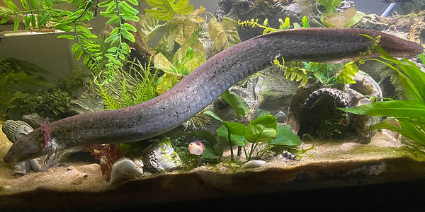
Murphy, a greater siren. Credit to @t0ost on Discord.
Amphibians and Other Ambystoma Species
"...Although genomic approaches would shed light on these unique traits and aid conservation, sequencing and assembly of amphibian genomes has lagged behind other taxa due to their comparatively large genome sizes. Fortunately, the development of long-read sequencing technologies and initiatives has led to a recent burst of new amphibian genome assemblies... Here, we analyse 51 publicly available amphibian genomes to evaluate their usefulness for functional genomics research. We report considerable variation in genome assembly quality and completeness and report some of the highest transposable element and repeat contents of any vertebrate. Additionally, we detected an association between transposable element content and climatic variables. Our analysis provides evidence of conserved genome synteny despite the long divergence times of this group, but we also highlight inconsistencies in chromosome naming and orientation across genome assemblies. We discuss sequencing gaps in the phylogeny and suggest key targets for future sequencing endeavours. Finally, we propose increased investment in amphibian genomics research to promote their conservation."
"In this overview, we briefly review certain features of mammalian immune systems (covered in much more detail in virtually any textbook of immunology), summarize the evidence that salamanders have a subdued immune system (based on functional evidence mostly from the works of Delanney, Cohen, Charlemagne, and Tournefier), describe the unpublished data that we and our collaborators have generated to show that the axolotl has both a Major Histocompatibility Complex (MHC) and functional T lymphocytes, and conclude with our present interpretation of why the axolotl and other urodeles have subdued immune responses."
"This talk covers aspects of amphibian anatomy and physiology with immediate relevance to the veterinarian dealing with amphibians."
"The amphibian skin is not only a coating; it consists of a system of activities related to respiration, osmosis, and thermoregulation and, because of that, enables the creature to survive on land. Besides that, the skin synthesises a broad array of bioactive compounds that act as defences against the environment and pathogenic agents. Still, they also play a wide range of other biological roles. Antioxidant peptides in the amphibian system can hinder lipid peroxidation and defend the body against free radical attacks. Thus, when the normal balance of oxidative stress is broken by excessive production of free radicals, the intake of sufficient amounts of antioxidant peptides may reinstate the imbalance, and, therefore, minimise the risk of developing a disease. However, this is the least developed field and the research on antioxidant amphibian peptides is particularly underrepresented in the literature. The current paper provides an overview of the structure and function of antioxidant peptides in amphibian skin, aiding in a better understanding of how particular species have evolved defences against environmental assault and the future development and utilisation of amphibian antioxidant peptides."
"...The axolotl, a neotenic salamander, has become the model organism for regeneration research. Great advances have been made providing a detailed understanding of the morphological and molecular processes involved in limb regeneration. However, it remains largely unknown how limb regeneration varies across salamanders and how factors like variable life histories, ecologies, and limb functions have influenced and shaped regenerative capacities throughout evolution... Gaining insights into variable aspects of regeneration under natural conditions and after conspecific biting rather than controlled amputations adds important new datapoints for understanding the evolutionary framework of regeneration and provides a broader context for interpreting findings made in the model organism axolotl."
"The major histocompatibility complex (MHC) is a cluster of functionally related genes encoding proteins which, among other functions, mediate immune system activation. While the MHC of many vertebrates has been extensively studied, less is known about the amphibian MHC. This represents an important knowledge gap because amphibians mark the evolutionary transition from an aquatic to a terrestrial lifestyle and often maintain a biphasic lifestyle. Hence, they tend to be exposed to both aquatic and terrestrial pathogen communities, providing opportunities to gain fundamental insights into how the immune system responds to different environmental challenges."
"Here we summarize the current knowledge of salamander limb development under consideration of the fossil record to provide a deep time perspective of this evolutionary pathway and highlight what data will be needed in the future to gain a better understanding of salamander limb development specifically and tetrapod limb development and evolution more broadly."
"We gathered current knowledge about the roles amphibians play in the wild and explained how their decline can impact ecosystems. We also highlighted how new technologies such as satellite remote sensing and geo-spatial sciences as well as citizen science can help track these changes and support conservation efforts. Importantly, we call for improved public awareness and educational programs, use of indigenous knowledge, and stronger policies that recognize amphibians not just as endangered species, but as vital parts of functioning ecosystems..."
"The composition and diversity of animal-associated microbial communities are shaped by multiple ecological and evolutionary processes acting at different spatial and temporal scales. Skin microbiomes are thought to be strongly influenced by the environment due to the direct interaction of the host’s skin with the external media. As expected, the diversity of amphibian skin microbiomes is shaped by climate and host sampling habitats, whereas phylogenetic effects appear to be weak. However, the relative strength of phylogenetic and environmental effects on salamander skin microbiomes remains poorly understood. Here, we analysed sequence data from 1164 adult salamanders of 44 species to characterise and compare the diversity and composition of skin bacteria. We assessed the relative contribution of climate, host sampling habitat, and host phylogeny to the observed patterns of bacterial diversity. We found that bacterial alpha diversity was mainly associated with host sampling habitat and climate, but that bacterial beta diversity was more strongly associated with host taxonomy and phylogeny. This phylogenetic effect predominantly occurred at intermediate levels of host divergence (0–50 Mya). Our results support the importance of environmental factors shaping the diversity of salamander skin microbiota, but also support host phylogenetic history as a major factor shaping these bacterial communities."
"...Caudates (salamanders) in the genus Ambystoma represent a challenge for rearing because of their aggressive nature, and several recent projects have found higher than normal stress levels in captive-reared salamanders. In this study I compared various captive-rearing scenarios designed to minimize stress levels in Spotted Salamanders (A. maculatum). I reared larvae in five treatments, three that varied in starting density (six, 12, and 30 larvae per 1000 L tank), one with high food, and one with added environmental complexity to the tanks (sticks and refugia). After metamorphosis, I weighed the salamanders and collected blood samples to determine the relative abundance of two white blood cell types (neutrophils and lymphocytes) to calculate neutrophil-lymphocyte (N-L) ratios, which covary with stress hormone levels… The treatment that led to the largest salamanders overall and the only one where N-L ratios were similar to those from the wild was the treatment with the lowest starting density (six larvae per 1000 L tank). Furthermore, N-L ratios across all treatments were a direct function of body size; larger salamanders exhibited lower stress."
"The aim and structure of this review are to guide the reader through the progress on the basic questions that regeneration biologists are asked time and again: why does amputation start regeneration? How does it know when to stop? How does the limb “know” which parts of the limb to regenerate? Are cells reprogrammed or dedifferentiated to regenerate? How did it evolve? An important recent synergy has been parallel progress in multiple invertebrate and vertebrate models. It is not possible to generally cover all systems here, but rather this review anchors itself on progress in salamander limb regeneration and refers to parallel or supporting information from other regeneration systems to fulfill illustrative aims."
"Amphibians are sensitive to contaminants, including NaCl, because of their porous skin and osmoregulatory adaptations to freshwater. However, similarities and differences between effects of wastewaters and road salts have not been investigated. Therefore, we investigated the relative influence of wastewaters and NaCl at equivalent concentrations of chloride on three larval amphibian species that occur in areas with increased salinity. We determined acute toxicity and growth effects on Boreal Chorus Frogs (Pseudacris maculata), Northern Leopard Frogs (Rana pipiens), and Barred Tiger Salamanders (Ambystoma mavortium). We posited that wastewaters would have additive effects on amphibians compared to NaCl because wastewaters often have additional toxic heavy metals and other contaminants. For NaCl, toxicity was higher for frogs than the salamander... The negative influence of salts on imperiled amphibians underscores the need to restore landscapes with increased salinity and reduce future salinization of freshwater ecosystems."
This catalogue defines the 29 species of Ambystoma salamanders found in the Americas around 1969. Physical descriptions, habitat distribution, fossil records, etymology, and relevant history are discussed.
Last updated on March 11 2026